Reference


OpenAI's Answer
Synovial osteochondromatosis is typically diagnosed using a variety of imaging techniques. These include:
-
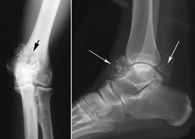
Plain Radiographs (X-rays): This is usually the first imaging technique used to diagnose synovial osteochondromatosis. Radiographs may reveal multiple intraarticular calcifications. They often depict numerous small, round, calcified bodies within the joint source.
-
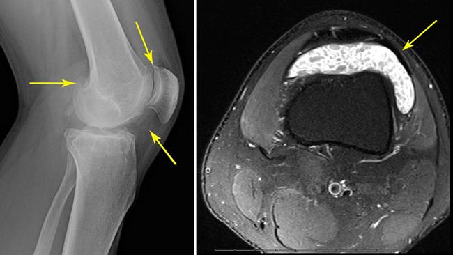
Magnetic Resonance Imaging (MRI): MRI is particularly useful in early stages of the disease, especially when calcifications are not yet visible on X-rays. It provides a detailed view of the joint and can show cartilage nodules and loose bodies within the joint source.
-
Computed Tomography (CT): CT scans can show the extent of mineralization in the joint and help in assessing the full extent of the disease, especially in cases where MRI is not conclusive source.
-
Ultrasound: This can be used to identify loose bodies within the joint and assess synovial proliferation. It is less commonly used but can be beneficial in certain cases source.
These imaging techniques together help to diagnose synovial osteochondromatosis, with MRI often being the most definitive method due to its ability to show early changes in the joint not visible on other modalities.
Here are some images that illustrate the condition:
For more detailed visual examples, you might want to refer to specialized radiology resources or an orthopedic text.
Follow Up
Related
What are the differential diagnoses for intra-articular bodies in synovial chondromatosis?
Why can't prion diseases be diagnosed solely based on symptoms?
What are the challenges in diagnosing prion diseases during the incubation period?
What significant medical condition was Dean Canto diagnosed with?
